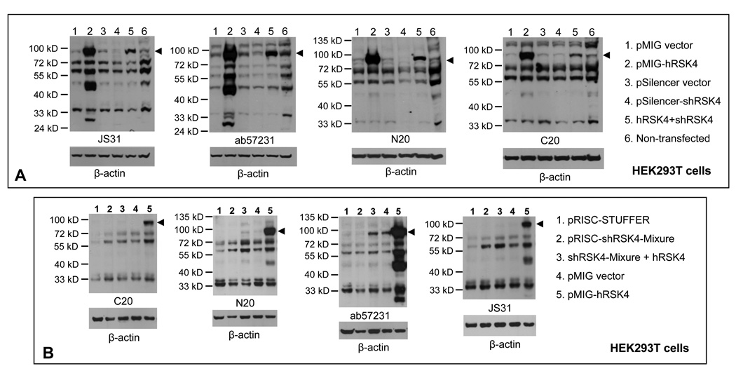
Fig. 7.
Verification of hRSK4 proteins with siRNA. A: HEK293T cells were transfected with an RSK4 shRNA in a pSilencer vector, alone or together with the pMIG-hRSK4 cDNA used in figure 5A. Controls include transfections with each empty vector and non-transfectants. Immunoblots with four different antibodies all show that the 90-kD protein (arrowhead) expressed from the hRSK4 cDNA is dramatically down regulated by the shRNA (lanes 2 vs 5), so as well the 48-kD protein. The 72- and 55-kD proteins are only partially decreased and the decrease is detected only by some of the four antibodies. B: HEK293T cells were transfected with the hSRK4 cDNA construct, alone or together with a mixture of five RSK4 shRNAs expressed from a pRISC-Stuffer vector.17 Controls include cells transfected with each empty vectors. Immunoblots with indicated antibodies again show that the 90-kD protein is dramatically down regulated by the shRNA cocktail whereas the 72- and 55-kD proteins are only partially decreased (lanes 3 vs 5). After blotting with an RSK4 antibody, each membrane was stripped and then re-blotted with β-actin antibody to show the protein loading.

