Abstract
Purpose
The aim of this study was to analyse the management of displaced paediatric supracondylar humerus fractures at our Level I Trauma Centre and to determine clinical and radiographic long-term results following operative treatment.
Methods
Clinical and radiological results of 78 paediatric patients (29 female, 49 male; mean age 5.1 years) with supracondylar humerus fractures, treated from 1992 to 2004, were evaluated. Gartland’s classification yielded 32 type II, 44 type III and further two flexion injuries. In all patients the follow-up period exceeded 12 months. Assessment after an average of 8.1 years (1.1–19.5) included neurovascular examination, Flynn’s criteria (elbow function and carrying angle), pain, complications (infections, growth disturbances or iatrogenic nerve injuries) and measurement of the humeroulnar angle.
Results
According to Flynn’s criteria 73 patients (93.5 %) had a satisfactory outcome, while five (6.4 %) were graded as unsatisfactory (two due to cubitus varus and three because of limited elbow motion). The visual analogue scale (VAS) score averaged 0 (range 0–1) and the mean carrying angle measured 8.4° (−8 to 20°), compared to 10.8° on the contralateral side (2–20°). Injury-related complications yielded absent pulses in four (5.1 %), five (6.4 %) primary median, two (2.6 %) primary radial and one (1.3 %) primary ulnar nerve injury. Treatment-related complications included a secondary displacement and one iatrogenic radial nerve palsy. Based on primary nerve lesion as a dependent variable, statistical analysis showed that age had a significant influence revealing that older paediatric patients had a significantly higher risk (p = 0.02). Functional outcome as a dependent variable revealed an indirect proportion to the clinical carrying angle, achieving statistical significance (p < 0.01).
Conclusions
Crossed pinning in paediatric supracondylar humerus fractures is an effective method. Evaluation of the outcome in our study group demonstrated good results with the treatment approach described.
Introduction
Supracondylar humerus fracture is one of the most common injuries in children accounting for seven to nine percent of all childhood fractures [1]. At the same time it is a troublesome injury with complications including neurovascular damage, compartment syndrome, Volkmann’s ischaemic contracture and malunion, especially cubitus varus [2–4]. Extension-type injuries are classified according to Gartland’s criteria as non-displaced fractures (type I), hinged fractures with the posterior cortex intact (type II) and completely displaced fractures (type III) [5]. In displaced fractures the treatment of choice is closed reduction and percutaneous pinning [2, 4, 6–8]. The biomechanical superiority of crossed K-wires was described by several authors [2, 4, 9]. Due to the risk of iatrogenic ulnar nerve lesions some authors prefer lateral pin placement [2, 8]. Others use a medial mini-open approach to visualise the ulnar nerve [6].
Thus, the aim of this study was to evaluate the management of displaced paediatric supracondylar humerus fractures with closed reduction followed by percutaneous crossed K-wire placement and open access in selected cases only (where closed reduction failed or due to neurovascular damage) and to determine the clinical and radiographic long-term results following operative treatment.
Methods
After approval of the Institutional Review Board for the recruitment of patients, we reviewed patient records at our Level I Trauma Centre from September 1992 to June 2004, matched for the following inclusion criteria: displaced supracondylar humerus fractures (Gartland type II and III injuries), with open epiphyseal plates of the distal humerus, with a minimum follow-up of one year. Skeletally mature adults and patients with previous or concomitant ipsilateral elbow fractures were excluded. This initial search yielded a total of 117 consecutive paediatric patients with displaced supracondylar humerus fractures. During the recruitment process for this study, it was found that ten patients had no contact details at all, nine patients had wrong contact details and 20 patients did not reply.
Finally, there were 78 patients (66.7 %) available for follow-up. In all patients the follow-up period exceeded 12 months, with an average of 8.1 years (range 1.1–19.5 years) after the injury. The mean age of the 78 patients (29 female, 49 male) was 5.1 years (range 1.6–10.7, deviation 2.3), the right elbow was injured in 30 and the left in 48 patients. Concomitant injuries included one ipsilateral distal radius fracture, two ipsilateral and one contralateral forearm fracture, one ipsilateral humerus fracture, three ipsilateral bicondylar humerus fractures and one patient suffered multiple injuries (including an intracerebral haematoma, a skull base fracture, an ipsilateral pneumothorax and humerus fracture and a contralateral clavicle fracture).
Fracture classification
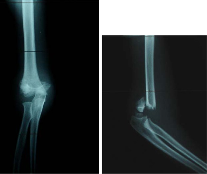
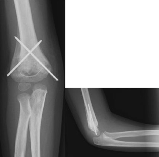
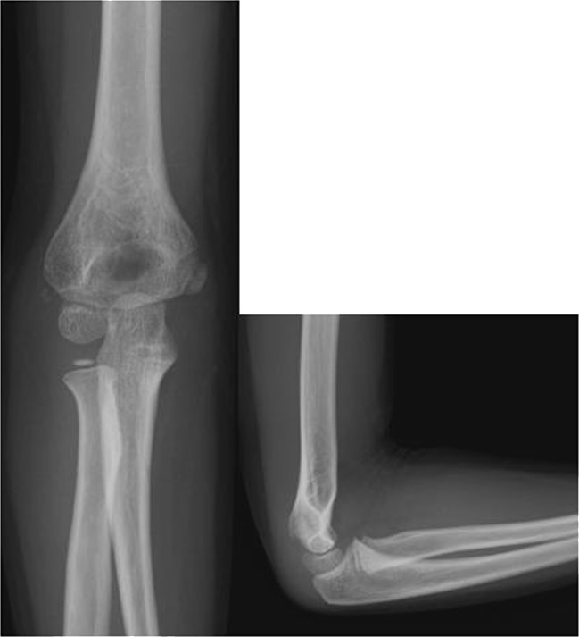
According to the classification system of Gartland, we had 32 patients with a type II injury and 44 patients with a type III injury (Figs. 1, 2, and 3). There were two flexion-type injuries in our series.
Fig. 1.

A 4-year-old girl with Gartland type III fracture
Fig. 2.
Four weeks postoperatively (same patient as Figure 1)
Fig. 3.
At follow up (two years postoperatively) (same patient as Figure 1)
Surgical technique
Under general or regional anaesthesia in a supine position on the operating table, closed reduction was performed under fluoroscopic guidance. With the elbow in hyperflexion one pin was inserted from the lateral side of the elbow across the lateral cortex engaging the medial cortex. Then the elbow was extended to less than a 90° position and the ulnar nerve was palpated. The medial pin was then placed beginning at the medial epicondyle to engage the lateral cortex in again a hyperflexed elbow position. If closed reduction failed or in cases of neurovascular damage (mini)-open reduction was performed and the described fixation technique was used subsequently.
Clinical and radiographic examination
Follow-up monitoring included regular clinical and radiographic examination of the patients at one, four, six and 12 weeks and 12 months after injury. The cast was removed at the four week follow-up appointment, while pin removal took place at the six week visit. All patients were followed up for at least one year after the initial treatment.
Clinical evaluation included neurovascular examination, measurement of the range of motion (ROM) of the injured elbow, assessment of the carrying angle [1], pain and determination of any complications such as infections, growth disturbances or iatrogenic nerve injuries. The ROM of the injured elbow was measured by a manual full-circle goniometer. The clinical results were graded according to Flynn’s criteria, which are based on the carrying angle and the elbow motion [10]. The carrying angle was measured by a full-circle goniometer and compared with that of the contralateral arm. Pain was assessed according to the visual analogue scale (VAS) that allowed for numeric responses on a scale from 0 (no pain) to 10 (worst pain) [11]. Radiographic evaluation included anteroposterior and lateral radiographs of the injured elbow, which were made intraoperatively and at each follow-up examination. The humeroulnar angle was calculated on the anteroposterior radiograph with the method of Webb and Sherman at the final follow-up examination [12].
The humeroulnar angle was defined by the intersection of the midhumeral line with the line drawn from the proximal midpoint of the ulna to the distal midpoint of the ulna in the anteroposterior view on a radiograph with the elbow extended to 0° and the forearm supinated [12].
Statistical analysis
For statistical analysis we performed a multiple regression analysis with a 95 % confidence interval. To determine statistical significance of corresponding variables (age, sex, open or closed reduction, Gartland type, clinical carrying angle, functional outcome and nerve injury) we used a p value <0.05. Functional outcome and incidence of a nerve lesion represented the dependent variables.
Results
During the study period 78 surgically treated paediatric patients with supracondylar humerus fractures met the criteria for inclusion and were finally enrolled in this series. Of these patients, 41 were treated by closed reduction and percutaneous crossed K-wire fixation, whereas 36 underwent open reduction due to soft tissue interposition or comminution. In one case open access was used due to suspected primary vascular damage.
After the surgical procedure, an anteriorly split long arm cast was applied with approximately 90° of elbow flexion and neutral forearm rotation.
Clinical outcome
According to Flynn’s criteria 73 patients (93.5 %) had a satisfactory outcome, while five (6.4 %) were graded as unsatisfactory. Two patients were graded as unsatisfactory due to cosmetic factors. One of these had a cubitus varus of −8° resulting in a difference of 18° in comparison to the uninjured side. In the other patient the clinical carrying angle was 0° and differed by 14° in comparison to the uninjured elbow. Both patients had regained unlimited elbow function at follow-up. In the remaining three patients classified as unsatisfactory, two had an extension deficit and one a flexion deficit of 20°. At the one year follow-up examination, none of the patients complained about any relevant pain symptoms. At this time the average VAS score was 0 (range 0–1). The mean carrying angle measured 8.4° (range −8 to 20°), compared to 10.8° on the contralateral uninjured side(range 2–20°).
Radiographic outcome
Successful fracture healing was achieved in all of our patients (100 %). One patient had signs of a delayed union, but at the six month follow-up examination, X-rays showed a stable osseous union. Incomplete primary reduction was not seen in any of our patients. Secondary displacement was noted in one patient following closed reduction and percutaneous pinning at the one week follow-up examination. This patient was reoperated and the fixation was performed with two lateral and one medial K-wire. Finally, two malunions were found in our series (cubitus varus). The humeroulnar angle averaged 10.1° (range −8 to 22°).
Complications
Injury-related complications were seen in 12 patients (15.4 %), including absent pulses in four patients (5.1 %), five (6.4 %) primary median nerve injuries, two (2.6 %) primary radial and one (1.3 %) primary ulnar nerve injury. The pulses were restored after closed reduction in all but one patient, where exploration of the brachial artery revealed kinking due to the proximal fracture fragment, but no laceration. Postoperative sonographic and clinical examination revealed normal pulses and a well-perfused hand. All but one median, radial and ulnar nerve palsies, which were present pre-operatively, were associated with the fracture and resolved spontaneously after an average of 5.5 months (range 0–104 weeks) (Table 1). In the remaining patient with radial nerve palsy revision surgery revealed compressive scar formation in the nerve surrounding soft tissue next to the fracture and decompression was performed. This patient recovered completely four weeks later (12 weeks after the initial trauma).
Table 1.
Complications
| Injury-related complications | Total no. of patients 12 (15.4 %) | Therapy | Resolved post-injury | Outcome | |
|---|---|---|---|---|---|
| Primary median nerve injury | 5 (6.4 %) | 1 | Expectant | After 0 weeks | Complete recovery |
| 2 | Expectant | After 2 weeks | Complete recovery | ||
| 3 | Expectant | After 6 weeks | Complete recovery | ||
| 4 | Physiotherapy | After 52 weeks | Complete recovery | ||
| 5 | Physiotherapy | After 104 weeks | Complete recovery | ||
| Primary radial nerve injury | 2 (2.6 %) | 1 | Expectant | After 4 weeks | Complete recovery |
| 2 | Revision surgery after 8 weeks | After 12 weeks | Complete recovery | ||
| Primary ulnar nerve injury | 1 (1.3 %) | 1 | Expectant | After 10 weeks | Complete recovery |
| Absent radial pulse | 4 (5.1 %) | 1 | Closed reduction | Immediately | Complete recovery |
| 2 | Closed reduction | Immediately | Complete recovery | ||
| 3 | Closed reduction | Immediately | Complete recovery | ||
| 4 | Exploration of brachial artery | Immediately | Complete recovery | ||
| Treatment-related complications | 2 (2.6 %) | ||||
| Iatrogenic radial nerve injury | 1 (1.3 %) | 1 | Expectant | After 13 weeks | Complete recovery |
| Iatrogenic ulnar nerve injury | 0 | ||||
| Iatrogenic median nerve injury | 0 | ||||
| Secondary displacement | 1 (1.3 %) | Reoperation (3 K-wires) | Flynn 1, no complaints |
Treatment-related complications were seen in two patients, as we noted a secondary displacement due to instability and one iatrogenic radial nerve palsy. During placement of the medial K-wire the wrist was noticed to jerk in dorsiflexion as the lateral humeral cortex was penetrated. The K-wire was drawn back to the lateral cortex of the humerus. Postoperatively, the power of wrist extension was significantly reduced in comparison to the contralateral side, but recovered completely without further intervention within 13 weeks. We did not see any deep infections after surgical treatment. Early pin removal due to migration or superficial infection was performed in five cases (6.4 %). There was no case of iatrogenic ulnar nerve injury
Statistical results
Based on primary nerve lesion as a dependent variable, statistical analysis showed that age had a significant influence on this variable revealing that older paediatric patients had a significantly higher risk (p = 0.02). With each increasing year of life there is a 3.5 % higher probability of sustaining a primary nerve injury. None of the other variables showed any significant influence.
With the functional outcome as a dependent variable, statistical results revealed an indirect proportion between the dependent variable and the clinical carrying angle, achieving statistical significance (p < 0.01). All of the other variables failed to reach statistical significance.
Discussion
The main goal in paediatric supracondylar fracture treatment is to safely achieve a stable reduction to prevent displacement of the distal fragment and postoperative deformity [1, 6, 13], which historically has been reported to be as high as 17 % [6]. Internal rotation of the distal fragment is the major predisposing factor to varus deformity and is necessary for coronal varus tilt to occur [1, 6, 14]. Cubitus varus is the most frequently reported complication throughout the literature [10, 12]. In contrast, Green et al. described no cases of malunion in their series of 65 patients treated by crossed pin placement [6]. Shim and Lee reported one patient with a cubitus varus deformity in their series of 63 patients (1.6 %) treated by cross-fixation with three K-wires. Flynn et al. described three of 72 patients (4.2 %) with a cosmetically unsatisfactory result due to loss in carrying angle; none of these patients had a significant loss of elbow function (one normal ROM and two lacked only 5° of normal ROM) [10]. In our series two of 78 patients (2.6 %) had a comparable varus deformity at final follow-up. Multiple regression analysis of our data confirmed the results described in the literature [10, 12] that the carrying angle has no influence on the functional outcome, indicating that patients with a cubitus varus had a good elbow function. Our statistical analysis even revealed an indirect correlation between the carrying angle and the elbow function.
Associated vascular complications are common with supracondylar fractures because of the vulnerable position of the neurovascular structures in relation to the fracture fragments and haemorrhage [10]. Flynn et al. described an incidence of 18 % of vascular complications in their series of 52 patients and stated that their hospital served as a referral centre for complicated fractures from rural areas and thus some patients had repeated manipulation or presented two days after injury, thus incurring a higher complication rate [10]. Concerning vascular complications in our series, all of our patients were handled as emergencies, as stated above, and perhaps because of the prompt treatment, long-term vascular complications were avoided. This finding accords with D’Ambrosia who compared six treatment methods in 74 patients [13].
With regards to our method of treatment, percutaneous crossed K-wires achieved a successful outcome with a low incidence of major complications. In the current literature, Shim and Lee reported 63 consecutive paediatric cases treated by closed reduction and percutaneous cross-fixation with three K-wires (two parallel inserted from the lateral side, followed by one from the medial side). There was no iatrogenic ulnar nerve palsy in their collective [15]. This finding was in good agreement with our study, as we did not have any iatrogenic ulnar nerve palsy either. A medial skin incision for ulnar nerve exploration was recommended by several authors [6, 15–19]. On the one hand Shim and Lee stated that this would prolong the operation and increase the risk of infection. On the other hand most cases of postoperative ulnar neuropraxia that have been explored have revealed direct iatrogenic injury from the medial pin, including direct penetration or laceration of the nerve or tacking down the nerve sheet in a non-anatomical position [6]. For instance, Green et al. used this technique and found one patient (1.5 %) with a postoperative ulnar nerve injury [6]. Kocher et al. compared percutaneous crossed versus lateral pin placement in 52 patients and found no iatrogenic nerve injury in either group [2]. Royce at al. treated 143 children with percutaneous crossed K-wires and described one radial (0.7 %) and three ulnar secondary (2.1 %) nerve palsies [18]. The radial nerve palsy resulted from the medially inserted pin after penetration of the lateral cortex [18]. In our study group we had one iatrogenic radial nerve injury that recovered within 13 weeks without further surgical intervention. Subluxation of the ulnar nerve still remains a problem. In our series all patients were operated within six hours of injury; thus, the swelling was maybe not that prominent and the ulnar nerve palpable in the ulnar groove.
Myositis ossificans is a rarely described complication [13, 20], mainly after open reduction, and did not occur in our group.
In summary, crossed pinning in paediatric supracondylar humerus fractures is a safe and effective method with a low incidence of complications. It is crucial to achieve adequate reduction and K-wire stabilisation, especially to avoid malrotation and tilting of the distal fragment in the coronal plane to obtain correct alignment.
In cases were the ulnar nerve is palpable in the ulnar groove, blind percutaneous crossed pin placement is safe in our opinion. If closed reduction fails or ulnar nerve subluxation cannot be excluded, a medial mini-open approach to visualise the nerve is certainly safer and should be preferred.
Acknowledgments
Conflict of interest
The authors declare that they have no conflict of interest.
Author contributions
All authors have participated sufficiently in this work concerning conception and design of this study, drafting the article, critical revision for important intellectual content and final approval. I.K-M. takes responsibility for the integrity of the work as a whole, from inception to finished article (irena.krusche-mandl@meduniwien.ac.at)
Contributor Information
Irena Krusche-Mandl, Phone: +43-676-4310142, FAX: +431-40400-5939, Email: irena.mandl@gmx.at.
Silke Aldrian, Email: silke.aldrian@meduniwien.ac.at.
Julia Köttstorfer, Email: julia.koettstorfer@meduniwien.ac.at.
Astrid Seis, Email: astrid.seis@meduniwien.ac.at.
Gerhild Thalhammer, Email: gerhild.thalhammer@meduniwienac.at.
Alexander Egkher, Email: alexander.egkher@meduniwien.ac.at.
References
- 1.Wilkins KE. Fractures and dislocations of the elbow region. In: Rockwood CA, Wilkins KE, King RE, editors. Fractures in children. Philadelphia: Lippincott; 1990. pp. 509–721. [Google Scholar]
- 2.Kocher MS, Kasser JR, Waters PM, Bae D, Snyder BD, Hresko MT, Hedequist D, Karlin L, Kim YJ, Murray MM, et al. Lateral entry compared with medial and lateral entry pin fixation for completely displaced supracondylar humeral fractures in children. A randomized clinical trial. J Bone Joint Surg Am. 2007;89:706–712. doi: 10.2106/JBJS.F.00379. [DOI] [PubMed] [Google Scholar]
- 3.Ramachandran M, Skaggs DL, Crawford HA, Eastwood DM, Lalonde FD, Vitale MG, Do TT, Kay RM. Delaying treatment of supracondylar fractures in children: has the pendulum swung too far? J Bone Joint Surg Br. 2008;90:1228–1233. doi: 10.1302/0301-620X.90B9.20728. [DOI] [PubMed] [Google Scholar]
- 4.Pirone AM, Graham HK, Krajbich JI. Management of displaced extension-type supracondylar fractures of the humerus in children. J Bone Joint Surg Am. 1988;70:641–650. [PubMed] [Google Scholar]
- 5.Gartland JJ. Management of supracondylar fractures of the humerus in children. Surg Gynecol Obstet. 1959;109:145–154. [PubMed] [Google Scholar]
- 6.Green DW, Widmann RF, Frank JS, Gardner MJ. Low incidence of ulnar nerve injury with crossed pin placement for pediatric supracondylar humerus fractures using a mini-open technique. J Orthop Trauma. 2005;19:158–163. doi: 10.1097/00005131-200503000-00002. [DOI] [PubMed] [Google Scholar]
- 7.Mehlman CT, Strub WM, Roy DR, Wall EJ, Crawford AH. The effect of surgical timing on the perioperative complications of treatment of supracondylar humeral fractures in children. J Bone Joint Surg Am. 2001;83-A:323–327. doi: 10.2106/00004623-200103000-00002. [DOI] [PubMed] [Google Scholar]
- 8.Skaggs DL, Hale JM, Bassett J, Kaminsky C, Kay RM, Tolo VT. Operative treatment of supracondylar fractures of the humerus in children. The consequences of pin placement. J Bone Joint Surg Am. 2001;83-A:735–740. [PubMed] [Google Scholar]
- 9.Zionts LE, McKellop HA, Hathaway R. Torsional strength of pin configurations used to fix supracondylar fractures of the humerus in children. J Bone Joint Surg Am. 1994;76:253–256. doi: 10.2106/00004623-199402000-00013. [DOI] [PubMed] [Google Scholar]
- 10.Flynn JC, Matthews JG, Benoit RL. Blind pinning of displaced supracondylar fractures of the humerus in children. Sixteen years’ experience with long-term follow-up. J Bone Joint Surg Am. 1974;56:263–272. [PubMed] [Google Scholar]
- 11.Breivik H, Borchgrevink PC, Allen SM, Rosseland LA, Romundstad L, Hals EK, Kvarstein G, Stubhaug A. Assessment of pain. Br J Anaesth. 2008;101:17–24. doi: 10.1093/bja/aen103. [DOI] [PubMed] [Google Scholar]
- 12.Webb AJ, Sherman FC. Supracondylar fractures of the humerus in children. J Pediatr Orthop. 1989;9:315–325. [PubMed] [Google Scholar]
- 13.D’Ambrosia RD. Supracondylar fractures of humerus–prevention of cubitus varus. J Bone Joint Surg Am. 1972;54:60–66. [PubMed] [Google Scholar]
- 14.Smith L. Deformity following supracondylar fractures of the humerus. J Bone Joint Surg Am. 1960;42-A:235–252. [PubMed] [Google Scholar]
- 15.Shim JS, Lee YS. Treatment of completely displaced supracondylar fracture of the humerus in children by cross-fixation with three Kirschner wires. J Pediatr Orthop. 2002;22:12–16. doi: 10.1097/00004694-200201000-00004. [DOI] [PubMed] [Google Scholar]
- 16.Lyons JP, Ashley E, Hoffer MM. Ulnar nerve palsies after percutaneous cross-pinning of supracondylar fractures in children’s elbows. J Pediatr Orthop. 1998;18:43–45. doi: 10.1097/00004694-199801000-00009. [DOI] [PubMed] [Google Scholar]
- 17.Mostafavi HR, Spero C. Crossed pin fixation of displaced supracondylar humerus fractures in children. Clin Orthop Relat Res. 2000;376:56–61. doi: 10.1097/00003086-200007000-00009. [DOI] [PubMed] [Google Scholar]
- 18.Royce RO, Dutkowsky JP, Kasser JR, Rand FR. Neurologic complications after K-wire fixation of supracondylar humerus fractures in children. J Pediatr Orthop. 1991;11:191–194. doi: 10.1097/01241398-199103000-00010. [DOI] [PubMed] [Google Scholar]
- 19.Taniguchi Y, Matsuzaki K, Tamaki T. Iatrogenic ulnar nerve injury after percutaneous cross-pinning of supracondylar fracture in a child. J Shoulder Elbow Surg. 2000;9:160–162. doi: 10.1016/S1058-2746(00)90048-X. [DOI] [PubMed] [Google Scholar]
- 20.Kekomäki M, Luoma R, Rikalainen H, Vilkki P. Operative reduction and fixation of a difficult supracondylar extension fracture of the humerus. J Pediatr Orthop. 1984;4:13–15. doi: 10.1097/01241398-198401000-00003. [DOI] [PubMed] [Google Scholar]


