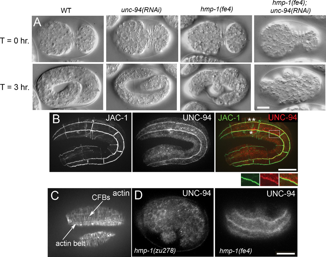
Figure 1. hmp-1(fe4);unc-94(RNAi) embryos arrest during embryonic elongation and epidermal UNC-94 localization to junctions requires hmp-1 function.
A. Nomarski images of representative embryos undergoing elongation are shown at the indicated time intervals. Embryos are initially oriented with anterior to the left and the ventral side up at T=0 hr., and lateral views are shown for all other time points. T=0 hr. shows embryos at the completion of enclosure. Wild-type (WT), unc-94(RNAi), hmp-1(fe4), and hmp-1(fe4); unc-94(RNAi) embryos are shown. No obvious defects in elongation are evident in the unc-94(RNAi) embryo. hmp-1(fe4) embryos develop mild body shape defects during elongation. 95% of hmp-1(fe4);unc-94(RNAi) embryos (n=93) elongate to the 1.5 fold stage or less, then retract to their original length. See Supplemental video S1, available online. B. Wild-type embryo expressing JAC-1::GFP to mark adherens junctions and stained with an affinity purified rabbit anti-UNC-94 antibody. Seam:ventral (*) and seam:dorsal (**) cell borders are indicated. UNC-94 is first detected at epidermal cell borders around the 2-fold stage of elongation. The color merge shows JAC-1::GFP in green and UNC-94 in red. Enlargement of the boxed region shows UNC-94 staining overlapping JAC-1::GFP. C. Wild-type stained with phalloidin. Circumferential filament bundles (CFBs) and the junctional actin belt are indicated. D. UNC-94 staining in wild-type and hmp-1 mutants. In wild-type elongation-stage embryos, 23% have UNC-94 localization at epidermal cell borders (n = 79). In hmp-1(zu278) embryos, 0% have UNC-94 localization at epidermal cell borders (n = 87). In hmp-1(fe4) embryos, 35% have UNC-94 localization at epidermal cell borders (n = 63). Note that this staining was done via freeze-cracking, which is known to disrupt actin, but preserves the UNC-94 epitope recognized by anti-UNC-94 antibodies. Disruption of junctional actin caused by this technique may account for the percentage of embryos exhibiting UNC-94 localization at epidermal cell borders. Bars = 10 µm.

