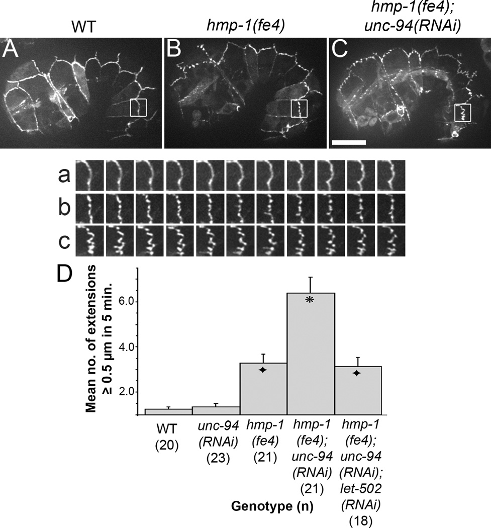
Figure 3. Adherens junctions of hmp-1(fe4);unc-94(RNAi) embryos exhibit abnormal dynamics.
(A, B, C) Images from spinning disk confocal movies of embryos expressing JAC-1::GFP are shown. a–c show an enlargement of the boxed regions in A – C at 30 second intervals for 5 minutes. Supplemental video S2, corresponding to a–c, is available online. In wild-type embryos, JAC-1::GFP localizes to epidermal cell borders; in hmp-1(fe4) embryos, there are occasional areas in which JAC-1::GFP is transiently extended away from its normal position. In hmp-1(fe4);unc-94(RNAi) embryos this behavior is more pronounced. Bar=10 µm. (D) Bar graph showing quantification of the number (mean ± SEM; n indicated in parentheses) of JAC-1::GFP extension longer than 5 µm formed at either the seam:dorsal or seam:ventral cell border during 5 minutes of filming. Embryos at comma to 1.5 fold stage were scored. Each extension was measured only once, at its longest length. Asterisk: significantly different from hmp-1(fe4) and hmp-1(fe4);unc-94(RNAi);let-502(RNAi) (Tukey test: p < 0.01). Black diamonds: not significantly different from hmp-1(fe4) (p > 0.5).

