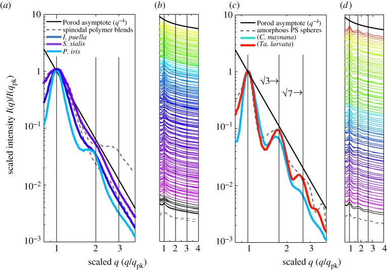
Figure 5.
SAXS structural diagnosis of amorphous photonic nanostructures in feather barbs. (a) and (c) depict, respectively, representative normalized azimuthal SAXS profiles for channel-type nanostructures from I. puella (Irenidae), S. sialis (Turdidae), and Pitta iris (Pittidae) and sphere-type nanostructures from C. maynana (Cotingidae) and Tangara larvata (Thraupidae) on a log–log scale exhibiting clearly distinguishable structural differences. The azimuthal profiles are scaled to compare across different colours and nanostructural sizes. (b) and (d) show, respectively, scaled azimuthal SAXS profiles for 159 channel- and 96 sphere-type nanostructures on a semi-log scale. The colour of each profile is coded to the approximate colour of the corresponding feathers based on its primary optical peak hue (pure UV colours shown in black). These azimuthal profiles are vertically displaced along the y-axis for clarity. In addition to the primary peak, the channel-type nanostructures (a,b) either have a weak to a pronounced shoulder at approximately twice the dominant spatial frequency, 2*qpk or lack any other significant feature, while the sphere-type nanostructures (c,d) exhibit one or more pronounced higher-order scattering peaks in addition to the primary peak at ratios of approximately √3 and √7 times qpk. The grey dashed lines in all figures plot the scaled experimental scattering profiles from two polymer mixtures undergoing spinodal decomposition [33,34] (a,b) and an amorphous film of self-assembled colloidal polymer spheres (c,d). The vertical lines at 1,2,3 (a,b) and at 1,√3 and √7 (c,d) are visual guides for the expected positional ratios for the SAXS peaks based on experimental observations of classical spinodal and nucleated, close-packed sphere morphologies, respectively.

