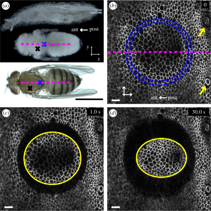
Figure 1.
Large-size annular severing in a fly dorsal thorax. (a) Developmental stages of a fruitfly Drosophila melanogaster. Top: larva. Middle: pupa, with pupal case removed (see electronic supplementary material, figure S1) and cuticle kept intact. Bottom: adult. Dashed lines represent the midline (symmetry axis). The x-axis is antero-posterior: anterior (head) towards the left, posterior (abdomen) towards the right. The y-axis is medio-lateral. Crosses: approximate positions of severing, along the midline in the scutellum (blue) and off-axis in the scutum (black). (b) Epithelial cell apical junctions marked by E-cadherin : GFP just before severing, t = 0−, here in an old pupa (see text for classification). Blue circles: two concentric circles define the annular severed region; the distance between circles corresponds to approximately one cell size. Yellow arrows: macrochaete used as spatial references to position the severed region. (c) First image after severing, t = 1 s. Yellow: fitted ellipse [35] (see §5). (d) Time t = 30 s after severing, showing a larger opening along y than x. Scale bars: (a) 1 mm, (b–d) 10 µm.

