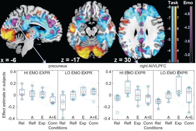
Figure 3.
Individual differences in the use of emotional words. Top: modulation of deactivations by individual differences in the tendency to use emotional words in the medial aspect of the cortex (left), in the orbitofrontal cortex (centre), and at the level of the angular gyrus (right), shown as parametric maps of t-values overlaid on a template T1-weighted brain. Activations and deactivations of the main reading task are displayed in yellow/orange and light blue, respectively, thresholded for display purposes at p = 0.005, uncorrected. In violet, the modulation by individual differences, at threshold p = 0.01, uncorrected, of the emotional contrast. The left side of the transversal slices is on the left. Bottom: box-plots of signal differences between conditions, displayed separately in subjects with high and low use of emotional words (“HI EMO EXPR”, “LO EMO EXPR”) at the follow-up post-scan recounting of the stories (for illustration purposes, the first and last thirds of a tertile split on the emotional words use scores were used). Here and in the following boxplots, data were centered. Data are divided according to textual description type, in the TCM terminology: Rel (relaxing) contains few emotional or abstract words, Refl (reflecting) is rich in abstract words (“A” on the x-axis), Exp (experiencing) is rich in emotional words (“E” on the x-axis), and Conn (connecting) contains both emotional and abstract words (“A + E” on the x-axis). The light blue ovals highlight the textual version with emotional words, which differ in the groups with high and low use of emotional words at recounting the scenes. AI/VLPFC: anterior insula/ventrolateral prefrontal cortex.

