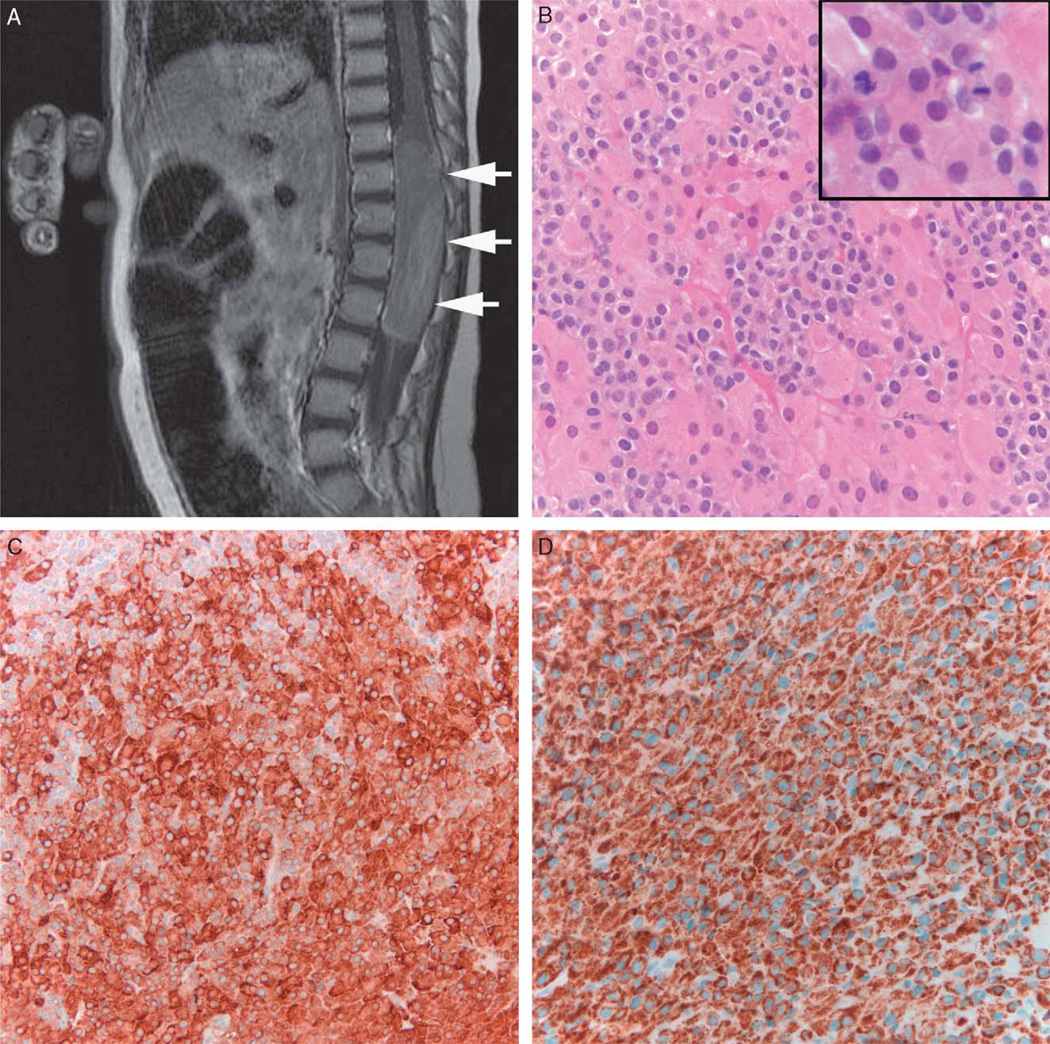
FIGURE 1.
Ectopic adrenocortical tumor. Sagittal T1 weighted MRI postcontrast demonstrates a large neoplasm in the lower spinal cord with mild to moderate heterogeneous enhancement (arrows) (A). The tumor histologically showed a biphasic cell population with large eosinophilic and small clear cells (B). Mitotic activity was brisk (inset). Immunohistochemical stains demonstrated strong reactivity for inhibin (C) and melan-A (D). MRI indicates magnetic resonance imaging.

