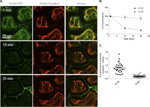
Figure 3. Perfusion and vascular permeability.
(A) The lamina propria of distal ileum was visualized via multiphoton, immediately following i.v. coinjection of 2 mg 10 kDa dextran-FITC (green) and 0.5 mg 70 kDa dextran-TR (orange) in saline. Time-lapse images were acquired every 30 s for up to 20 min following treatment of dextran dyes (Supplemental Video 1). (B) Quantitative kinetic analysis of the relative intravascular fluorescence up to 20 min, normalized to total fluorescence of each dye at 1 min postinjection. (C) Ratiometric analysis of relative fluorescence intensity of extra- versus intravascular dextran at 1 min postinjection; n = 3.

