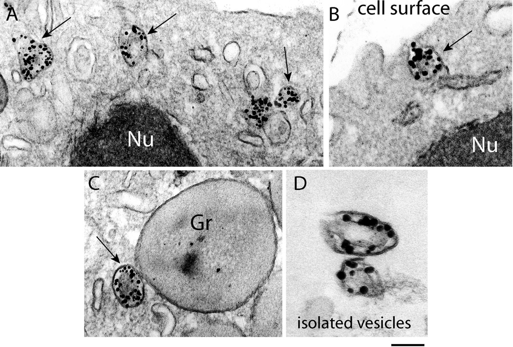
Fig. 4.
Large tubular carriers actively transport major basic protein (MBP). A–C. Eosinophil sombrero vesicles - EoSVs - (arrows) are observed in the cytoplasm by transmission electron microscopy (TEM) after immunonanogold labeling for MBP. Vesicles are seen beneath the plasma membrane in the cytoplasm (A), fused with the plasma membrane (B) and attached to an enlarged partially empty granule (C), typically indicative of PMD. D. EoSVs, isolated by subcellular fractionation, are densely labeled for MBP. Note that MBP is preferentially localized within the vesicle lumen. Eosinophils were stimuated by eotaxin as described in Material and Methods. Gr, specific granules; Nu, nucleus. Reprinted from (Melo et al., 2009) with permission. Scale bars: A, 400 nm; B, 230 nm; C, 250 nm; D, 200 nm.

