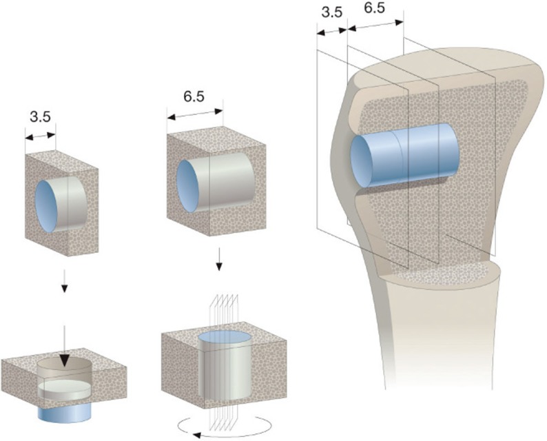
Figure 1.
Sectioning technique. Implant in situ in the tibia metaphyseal bone is illustrated to the right. Inner part of 6.5 mm was used for histomorphometry using the vertical-section method applying 4 sections around implant center after random rotation around implant axis (center panel). Outer part of 3.5 mm was used for mechanical testing (left panel).

