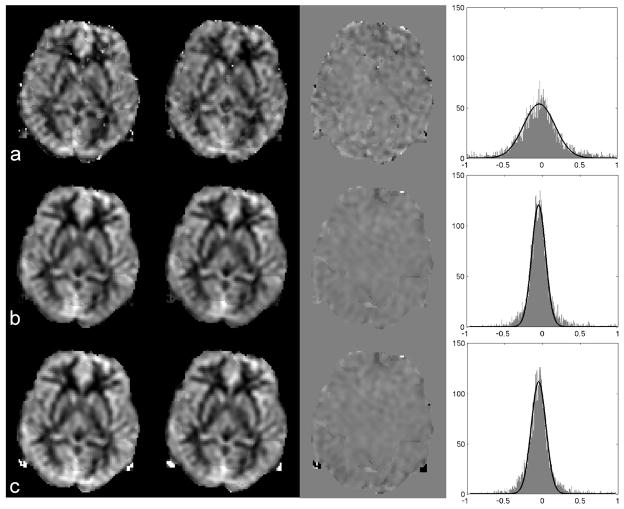
Fig. 4.
The perfusion maps generated from two repeated scans (the first two columns), the difference map between the two scans (the third column) and the distributions for the fractional change of perfusion between the two scans (the fourth column) using (a) Hadamard encoded method, (b) standard CASL with 1.5 s post-labeling delay (inferred from the long-labeling acquisition and an ATD map), and (c) PILOTS method. The fractional change of perfusion between two repeated scans using the standard method and PILOTS method has a narrower distribution than the fractional change of perfusion using the high-resolution Hadamard method, which indicates that the perfusion measurement using the standard and PILOTS method is more reproducible than that using the Hadamard encoded method.

