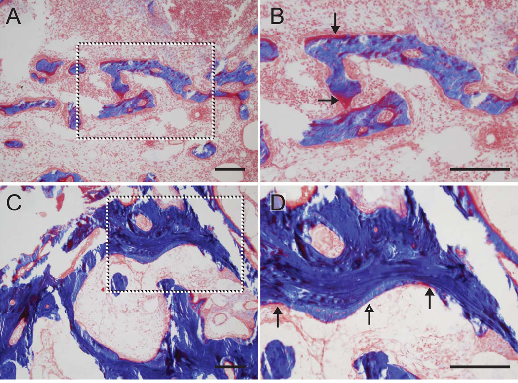
Figure 5.
New bone formation was evident in the trabeculae of the tunnel wall adjacent to the tendon in COL (A and B) and BMP-2/COL samples (C and D). The dotted boxes in the left panels are magnified and shown in the right panels. Osteoid can be seen (red stain, black arrowheads) in carrier control (COL) samples (A and B). Osteoid can be seen at the leading edges (red stain, black arrowheads) of newly mineralized bone (light blue stain, white arrowheads) in BMP-2/COL samples (C and D). There was more newly mineralized bone in the BMP-2 samples compared to controls. [COL: collagen carrier, 200um scale bars; Masson’s trichrome]

