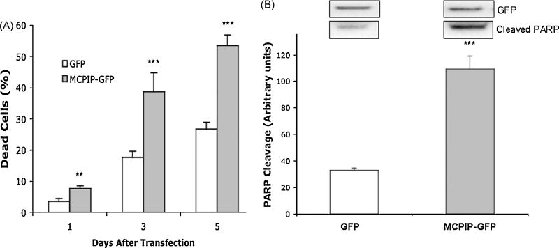
Figure 1. MCPIP increases cell death in NT2 cells.
(A) Percent cell death after MCPIP-GFP or GFP (Control) transfection. Cells were harvested at 1, 3, 5 days post transfection. Cell death was detected by trypan blue. (B) Immunoblot analysis of GFP expression and PARP cleavage in NT2 cells transfected with MCPIP-GFP or GFP. Analyzed 5 days post transfection. Protein values were normalized to Beta Actin expression. GFP expression demonstrates equal transfection efficiency. Note: MCPIP-GFP was approximately 90kDa compared to GFP alone (27 kDa). * Indicates statistical significance (**P<0.01, ***P<0.001).

