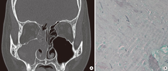
Fig. 1.
Diagnosis of allergic fungal sinusitis in the patient. (A) A non-enhanced computed tomography scan of the ostiomeatal unit (coronal view) reveals soft-tissue opacification in the right frontal, ethmoid, and maxillary sinuses. Arrow indicates allergic mucin, which filled the sinuses. (B) Staining of the allergic mucin with methenamine silver shows a few scattered fungal hyphae within the allergic mucin (methenamine silver stain,×400).

