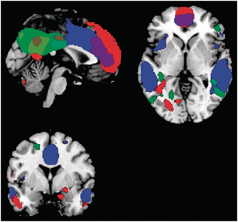
Fig. 1.
The three networks identified, the whole network is shown irrespective of group. All main areas of the default mode network are visible, including the anterior cingulate, posterior cingulate, medial middle/MTG, prefrontal cortex, precuneus and IPL. The anterior component is indicated in red, the middle DMN component in blue and the posterior component in green.

