Fig. 1.

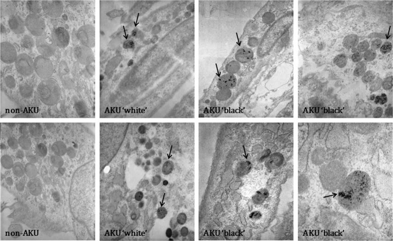
Deposition of ochronotic pigments, detected by TEM, in “white” and “black” AKU chondrocytes and their controls (non-AKU). Arrows indicate the presence of ochronotic pigments. Only representative images are shown.

Deposition of ochronotic pigments, detected by TEM, in “white” and “black” AKU chondrocytes and their controls (non-AKU). Arrows indicate the presence of ochronotic pigments. Only representative images are shown.