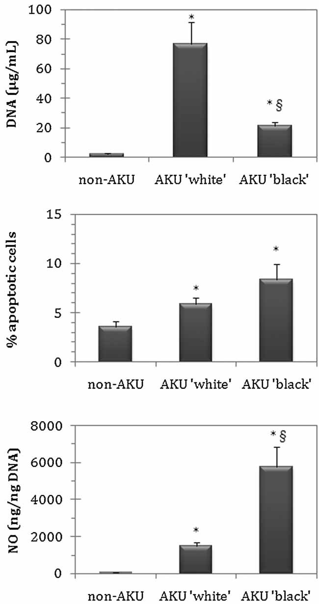
Fig. 2.

Cell proliferation (A), apoptosis (B), and NO release (C) in “white” and “black” AKU chondrocytes and their control (non-AKU). Cell proliferation was assayed by measuring the DNA content of cell pellets; apoptosis was assayed by Annexin V-FITC/propidium iodide staining and flow cytometry; NO release in culture supernatants was assayed by Griess reagent, as detailed under Materials and Methods Section. Experiments were performed in triplicate; data are presented as average values with standard deviation. Statistical significance compared to non-AKU control (*P ≤ 0.05) and between “white” and “black” AKU chondrocytes (§P ≤ 0.05) is indicated.
