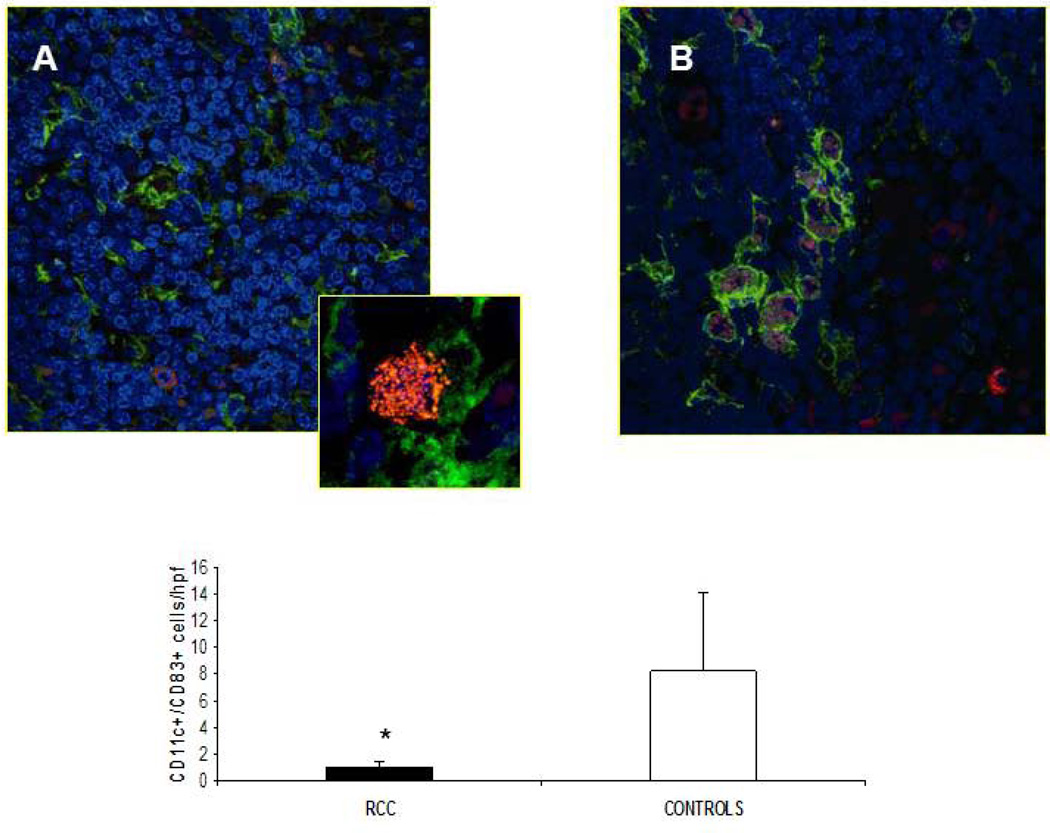
Fig. 5. Infiltrating CD11c+ CD83+ DCs in lymph nodes of RCC.
(A, B). The presence of mature DC was analyzed by confocal microscopy using antibodies that recognize mDC (anti-CD11c, green) and mature DC (anti-CD83, red), in RCC patients and healthy controls respectively (magnification 63×). Panel A shows a morphological particular of CD11c+/CD83+ DC (zoom 2×) (C). Quantification of CD11c-CD83 double-positive T cells. Results are expressed as mean ± SD of CD11c+ CD83+ in RCC patients versus healthy controls. (* P < 0.03).

