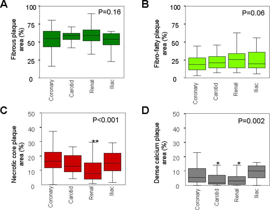
Figure 2.
Plaque composition at the smallest lumen site in four arteries. Box-whisker plots displayed the percent area of (A) fibrous plaque, (B) fibro-fatty plaque, (C) necrotic core, and (D) dense calcium of the whole plaque area. Renal arteries had less percentage of necrotic core (**p<0.001 vs. coronary). Carotid (*p=0.04 vs. coronary) and renal arteries (*p=0.01 vs. coronary) had less percentage of dense calcium.

