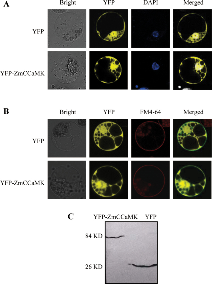
Fig. 2.
Subcellular localization of ZmCCaMK in maize protoplasts. (A, B) Protoplasts were isolated from the leaves of maize, and then were transfected with constructs carrying 35S:YFP-ZmCCaMK or 35S:YFP by PEG–calcium-mediated transformation. Fluorescence micrographs were taken after 16h of incubation by a laser confocal microscope. The nucleus was stained with DAPI dye (blue, A). The plasma membrane was labelled with FM4-64 steryl dye (red, B). Experiments were repeated at least five times with similar results. (C) Western blot analysis for YFP–ZmCCaMK fusion proteins with an anti-YFP antibody. Experiments were repeated at least five times with similar results. (This figure is available in colour at JXB online.)

