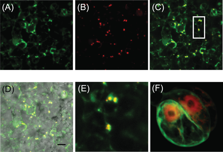
Fig. 4.
In vivo subcellular localization of PhAAE in MD petals and Arabidopsis protoplasts. (A) 35S:PhAAE-GFP and (B) the mCherry peroxisomal marker px-rk (Nelson et al., 2007) were co-infiltrated using A. tumefaciens in MD petal tissue. (C) The merged image of(A) and (B). (D) A merge of (C) and a brightfield image. (E) Magnification of the boxed area in (C). (F) Co-expression of a 35S:mCherry and 35S:PhAAE:GFP in Arabidopsis protoplasts. Representative images are shown. The scale bar in (D) represents 10 µm.

