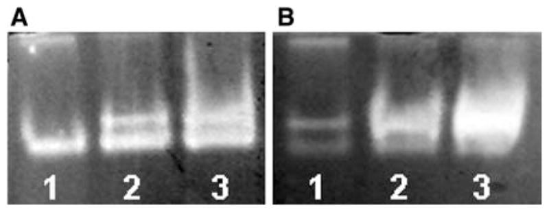
Fig. 6.

SOD activity in P. guilliermondii wild-type strain (A) and Δyfh1 mutant (B) correspondingly. Gels were stained for SOD activity following electrophoresis under non-denaturing conditions as described in the M&M section. Each lane was loaded with 0.04 mg of protein of cell free extract. Cells from early (1), middle (2), and late (3) exponential growth phase were used for the analysis
