Figure 1.
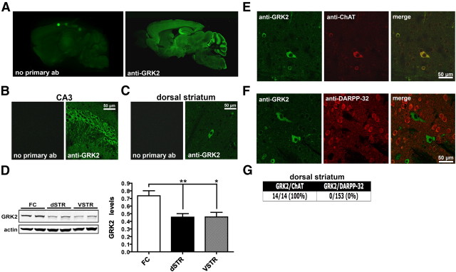
GRK2 expression in mouse brain. A–C, Brain sections prepared from C57BL/6J mice were immunostained either with a GRK2-specific antibody (anti-GRK2) or with the secondary antibody alone. Representative confocal images of GRK2 immunoreactivity throughout the brain (A), in the CA3 region of the hippocampus (B), or in the dorsal striatum (C) are shown (n = 5 mice). D, Western analyses of GRK2 levels in the FC, dorsal striatum (dSTR), and ventral striatum (VSTR) of C57BL/6J mice. Representative Western blots and the corresponding densitometric analyses are shown. GRK2 levels were normalized to actin in each respective lane. Data are mean + SEM; n = 6 mice for each group. **p < 0.01 and *p < 0.05 by unpaired t test. E, Immunofluorescence of dorsal striatal sections prepared from C57BL/6J mice that were costained with antibodies directed against either GRK2 (green) and ChAT (red) (E) or against GRK2 (green) and DARPP-32 (red) (F). A merge of both fluorescent channels in E and F is shown in the third column. G, Quantification of the number of ChAT- or DARPP-32-positive cells that were also found to be positive for GRK2 in dorsal striatal sections.

