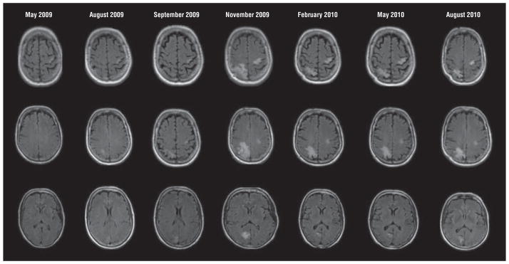
Figure 3.
Evolution of progressive multifocal leukoencephalopathy in the setting of rituximab therapy. The earliest lesion on fluid-attenuated inversion recovery images is seen in the May 2009 series, with slow evolution particularly in the right occipital lesion over the months from initial symptoms until diagnosis in September 2009 at the time of a brain biopsy. Plasma exchange was performed prior to the November 2009 series and the marked inflammatory response that followed then waned in this surviving patient. Typical of progressive multifocal leukoencephalopathy immune reconstitution inflammatory syndrome, the magnetic resonance images show marked expansion with perilesional edema that then resolved over months following immune control of progressive multifocal leukoencephalopathy. These same lesions demonstrated gadolinium contrast enhancement (not shown).

