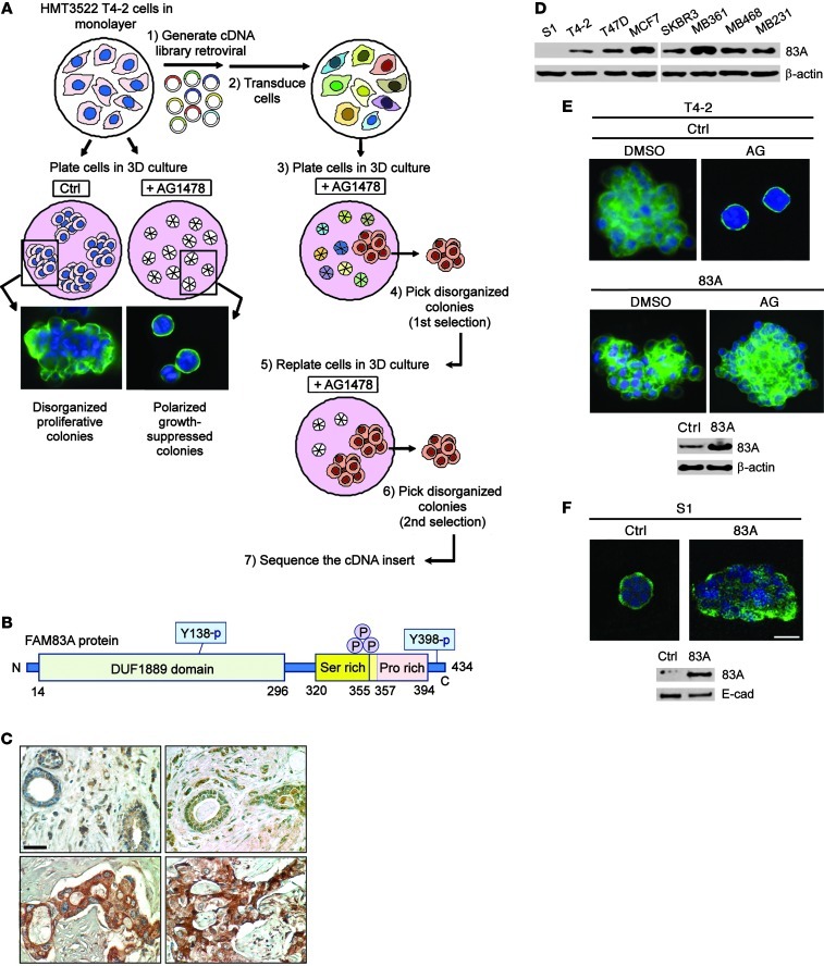
Figure 1. Discovery of FAM83A, a protein upregulated in breast cancer that confers resistance to EGFR-TKI AG1478.
(A) Process for the screen to identify FAM83A. Treatment of T4-2 cells with a small-molecule EGFR inhibitor, AG1478, induced reversion to a polarized acinar phenotype in 3D lrECM culture, as detected by α6 integrin staining (green) to delineate basal polarity (left); after transduction with the cDNA library, a subpopulation of cells disorganized in 3D culture after AG1478 treatment was isolated, expanded, and searched for the cDNA insert (right). (B) Protein domain structure of FAM83A. (C) Immunohistochemical analysis of FAM83A expression in normal (top) and malignant (bottom) breast tissue specimens. Whereas 0 of 16 normal cells were strongly positive for FAM83A staining, 45 of 48 malignant cells (94%) were positive. Scale bar: 50 μm. (D) FAM83A (83A) protein expression in a panel of breast cancer cell lines. (E and F) Effect of FAM83A overexpression in inhibiting AG1478-induced (AG) reversion of T4-2 cells (E) and ablating the polarized structure of S1 cells (F). Generation of T4-2 and S1 cell lines overexpressing FAM83A was confirmed by Western analyses. Scale bars: 50 μm.

