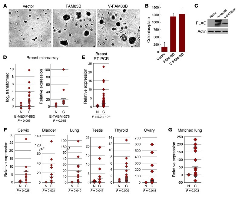
Figure 2. FAM83B drives AIG in HME1 cells and is elevated in human cancer specimens.
(A and B) HME1 cells were infected with a retrovirus encoding full-length FAM83B, VBIM-FAM83B, or a control retrovirus and analyzed for AIG. Original magnification, ×10. (C) Western analysis for FAM83B expression. (D) Microarray data sets E-MEXP-882 (4 normal, 18 cancerous) and E-TABM-276 (4 normal, 13 cancerous), acquired from Array Express, were used to examine transcriptional changes between normal (N) and cancerous (C) human breast tissues. (E and F) Origene TissueScan Cancer Panels (48-well breast cancer panels and a 384-well multi-cancer survey panel) were analyzed by real-time PCR for FAM83B. Shown is relative FAM83B expression for specimens that had a statistically significant difference between cancerous and associated normal tissues. n = 2 (normal bladder), 3 (normal thyroid and normal ovary), 4 (normal lung and normal cervix), 6 (normal testis), 9 (cancerous cervix), 16 (normal breast), 18 (cancerous thyroid), 19 (cancerous lung and cancerous testis), 21 (cancerous ovary), 22 (cancerous bladder), 191 (cancerous breast). (G) Relative expression of FAM83B in a panel of matched lung cancer and associated normal lung tissues. Each normal tissue was set equal to 1.

