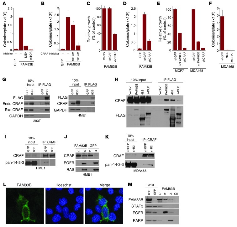
Figure 7. FAM83B-mediated transformation requires CRAF and mTOR signaling.
(A and B) FAM83B-expressing HME1 cells were treated with U0126 (1.5 μM), rapamycin (11 nM), or RAF kinase inhibitor I (100 and 600 nM) and assessed for AIG. (C and D) HME1 cells expressing FAM83B were infected with lentiviruses encoding shRNA targeting GFP or CRAF and assessed for growth and AIG. (E) MCF7 and MDA468 cells expressing shRNA targeting GFP, CRAF, or FAM83B were assessed for growth. (F) MDA468 cells expressing shRNA targeting GFP, CRAF, or FAM83B were assessed for AIG. (G) 293T cells were transfected with expression constructs encoding CRAF, GFP, and FAM83B, and immunoprecipitation was performed. Immunoprecipitation was also performed on lysates from HME1 cells stably expressing GFP or FAM83B. (H) 293T cells were transfected with expression constructs encoding CRAF, GFP, full-length FAM83B, FAM83B-482, and FAM83B-ΔDUF1669, and immunoprecipitation was performed. (I) Immunoprecipitation was performed on lysates from HME1 cells stably expressing GFP or FAM83B. (J) Cytoplasmic (C) and membrane (M) fractions from HME1 cells expressing GFP or FAM83B were analyzed by Western blotting. (K) Immunoprecipitation was performed on lysates from MDA468 cells expressing shRNA targeting GFP or FAM83B. (L) Immunofluorescence using confocal microscopy was performed in 293T cells transfected with GFP or FAM83B. Original magnification, ×63. (M) Whole cell extract (WCE), cytoplasmic (C), membrane (M), nuclear (N), and chromatin-bound (CB) fractions from 293T cells expressing GFP or FAM83B were analyzed by Western blotting. All experiments were performed in triplicate, and mean ± SD are shown. Lanes in I were run on the same gel but were noncontiguous (white lines).

