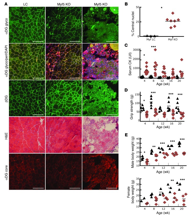
Figure 4. Mice with skeletal muscle Fktn deletion initiated at E8 show moderate to severe muscular dystrophy.
(A) Histology of iliopsoas muscle from 20-week-old myf5-Cre LC and KO mice provides evidence of moderate to severe dystrophy. Patchy or absent staining with αDG glyco-antibody while βDG and αDG core protein are still present indicates abnormal αDG glycosylation. Original magnification, ×20; scale bars: 100 μm. Note: Peripheral nerve twigs are still αDG glyco positive, confirming specificity of Fktn KO in muscle (e.g., asterisk). (B) Iliopsoas fibers with central nucleation in individual mice at 20 weeks of age. *P = 0.017; Mann Whitney test. (C) CK activity is elevated in KO mice at young ages. *P = 0.01–0.05, 4-week Myf LC versus KO; ***P < 0.001, 8-week Myf LC versus KO; Bonferroni test. (D) Forelimb grip strength (average of 5 pulls) is plotted according to age (*P = 0.01–0.05, 4-week Myf LC versus KO; ***P < 0.001, 8-, 16-, and 20-week Myf LC versus KO pairs; Bonferroni test. (E) Body weights of male and female mice at various ages. *P = 0.01–0.05, 4-week Myf LC versus KO M; **P = 0.001–0.01, 8-week Myf LC versus KO male and 16-week Myf LC versus KO female; ***P < 0.001, 12-, 16-, and 20-week Myf LC versus KO paired male and 20-week Myf LC versus KO female; Bonferonni test. Each data point represents 1 mouse; group means are shown. Black triangles, myf5-Cre LC; red diamonds, myf5-Cre Fktn-KO. Statistics were calculated for LC versus KO mice at each age.

