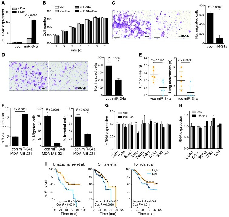
Figure 4. miR-34a regulates multiple biological properties of tumor cells.
(A–D) 344SQ_miR-34a cells and 344SQ_vector cells were cultured in the presence or absence of doxycycline (Dox). (A) Q-PCR analysis of miR-34a levels. (B) Cell numbers in monolayer. Migrating (C) and invading (D) cells in Boyden chambers were photographed and counted. Scale bars: 100 μm. (E) Primary tumor weight and total lung metastases from flank tumors in syngeneic mice (mean ± SD, n = 5). P values were determined by 2-tailed Student’s t test. (F) MDA-MB-231 cells were transiently transfected with a random sequence miR precursor molecule control or with pre–miR-34a precursor. Shown are Q-PCR analysis of miR-34a levels, expressed relative to control transfectants (set at 1.0), and migration and invasion assays in Boyden chambers. (G and H) Q-PCR analysis of epithelial (Cdh1 and Scrib) and mesenchymal (Cdh2 and Vim) markers and their transcriptional regulators (Zeb1, Zeb2, Snai1, Snai2, and Twist1) in 344SQ_vector and 344SQ_miR-34a cells (G) and in MDA-MB-231 cells transiently transfected with pre-miR control or pre–miR-34a precursor (H). Results are expressed relative to control transfectants (set at 1.0). Data are mean ± SD (n = 3). *P < 0.01. (I) Kaplan-Meier analysis of 3 independent cohorts of lung cancer patients (33–35), comparing the differences in risk between tumors with high (>0) or low (<0) scores (36), reflecting the presence or absence, respectively, of overlap with the murine miR-34a signature. P values from log-rank (differences between arms) and univariate Cox (gene signature score as a continuous variable) tests are shown.

