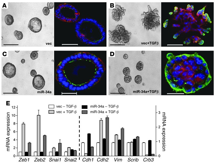
Figure 5. miR-34a blocks invasion, but does not reverse EMT.
(A–D) miR-34a repressed TGF-β–induced invasion in 3D Matrigel cultures. 344SQ_vector cells formed polarized epithelial spheres (A) that became hyperproliferative and invasive in the presence of TGF-β (B). 344SQ_miR-34a cells formed polarized epithelial spheres (C) that did not become invasive in the presence of TGF-β (D). Shown are light (left) and fluorescent (right) microscopic images of structures formed after 10 days in Matrigel containing doxycycline in the presence or absence of TGF-β (10 ng/ml). Blue, Topro-3; red, anti–α6 integrin; green, anti–ZO-1. Scale bars: 200 μm (light); 50 μm (fluorescent). (E) miR-34a did not abrogate TGF-β–induced EMT. Q-PCR analysis of epithelial markers (Cdh1, Scrib, and Crb3) and mesenchymal markers (Cdh2 and Vim) and their transcriptional regulators (Zeb1, Zeb2, Snai1, and Snai2) in 344SQ_vector and 344SQ_miR-34a cells after 10 days in Matrigel cultures containing doxycycline in the presence or absence of TGF-β. Results are expressed relative to empty vector transfectants treated without TGF-β (set at 1.0). Data are mean ± SD (n = 3).

