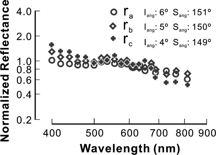
Figure 6. .
RNFL reflectance spectra along one bundle in a glaucomatous retina with normal-looking cytoskeleton. The spectra are different along the bundle. The slope of reflectance decrease at short wavelengths becomes shallower as the bundle area approaches the ONH. The whole spectrum looks flatter at ra, while the spectrum at rc is similar to that of normal. ra = 0.22 mm, rb = 0.33 mm, and rc = 0.44 mm from the center of the ONH. Reflectance spectra are normalized to the average reflectance at 500 to 560 nm and plotted on log-log coordinates. Iang, Sang: incident and scattering angles.

