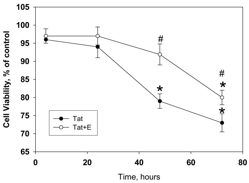
Figure 1. Estrogen treatment delays HIV-1 Tat 1-86 mediated decrease of cell viability in primary rat cortical cultures.
The graph demonstrates the decrease in live cells in primary cultures following exposure to 50 nM Tat 1-86. Addition of 10nM 17β-estradiol 24h prior to Tat 1-86 exposure was able to significantly delay onset of cell death in primary neuronal cultures. Data presented as mean values, n of sister cultures analyzed 5–10 per each time point. *P<0.05 as compared to non-treated controls, #P<0.05 as compared to Tat-treated vs. Tat+E treated cultures

