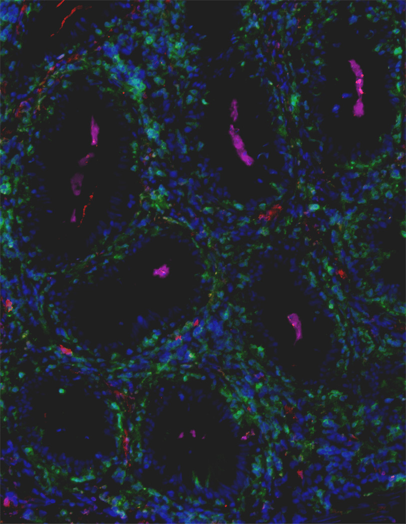
Figure 1. Extensive accumulations of IgT+ B cells are observed in the gut lamina propria and epithelium of rainbow trout surviving infection with the parasite Ceratomyxa Shasta.
Immunofluorescence staining of a gut cryosection from rainbow trout, three month post-infection with C. Shasta. Cryosection was stained for IgM (red), IgT (green) and C. Shasta (Magenta); nuclei are stained with DAPI (blue). Parasites (indicated by pink arrows) are localised within the gut lumen (dark area).

