Figure 2.

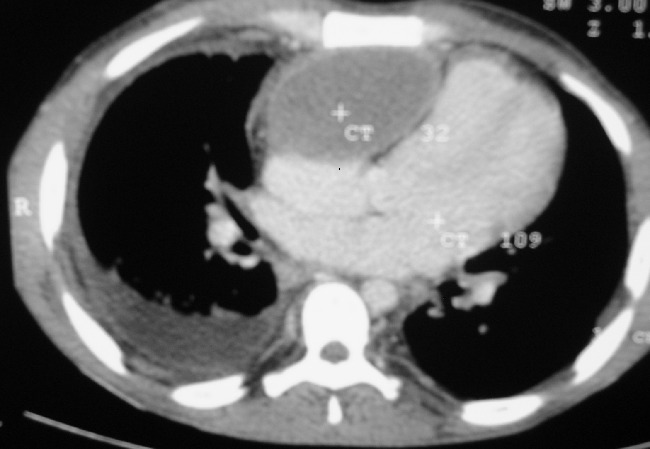
Axial CT image showing a retrosternal, anterior mediastinal fluid filled mass compressing the right ventricle and atrium. The mass has a well defined margin, non contrast enhancing and pushes RV to the left

Axial CT image showing a retrosternal, anterior mediastinal fluid filled mass compressing the right ventricle and atrium. The mass has a well defined margin, non contrast enhancing and pushes RV to the left