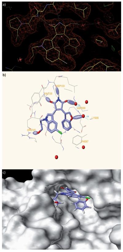
Figure 2.
X-ray co-crystal structure of benzofuran-3-yl-(indol-3-yl)maleimide bound to the GSK-3β (PDB ID: 3SD0; http://www.pdb.org). a) Electron density surrounding inhibitor 2 bound to the active site of GSK-3β. The Fo–Fc Fomit map is contoured at + 3s. b) Hydrogen bonds formed between GSK-3β and 2 are shown as dashed ellipsoids. The bromine atom of 2 is colored green. c) Solvent-exposed surface map of GSK-3β (grey) showing binding pocket for 2 and solvent exposure of the hydroxypropyl group.

