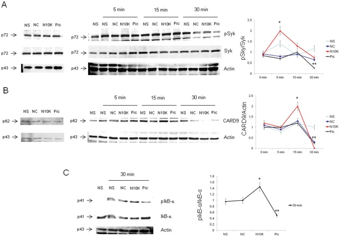
Figure 6. Dectin-1 signalling pathway activation.
PBMC (3×10⁶/ml) were pre-treated for 30 min in the presence/absence (NS) of NC, N10K (both 10 µg/ml) or piceatannol (Pic) (100 µM) and subsequently incubated for different times in the presence/absence of zymosan (50 µg/ml). After incubation, cell lysates were subjected to western blotting. Membranes were incubated with Abs to pSyk, Syk, CARD9, Actin, pIkB-α and IkB-α. Western blotting bands and protein normalizations are shown (A, B and C). *, P<0.05 (n = 5; N10K treated vs untreated cells). **, P<0.05 (n = 5; Pic treated vs untreated cells). Error bars denote s.e.m.

