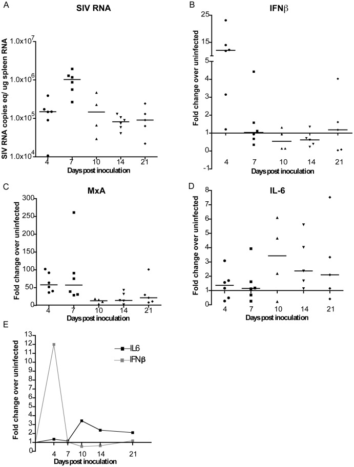
Figure 7. Characterization of the spleen in SIV-infected macaques during acute and early infection.
RNA isolated from the spleen of uninfected and infected macaques euthanized at 4, 7, 10, 14, or 21 days postinoculation (p.i.) was used to quantitate (A) SIV RNA copies equivalents/ug spleen RNA as well as mRNA expression of (B) IFNβ, (C) MxA, and (D) IL-6 by quantitative RT-PCR. mRNA expression is represented as fold change over the average of three uninfected spleen mRNA, calculated by ΔΔCt. Black bars indicate medians for each time point. (E) Summarizes the medians of IFNβ and IL-6 mRNA levels shown in (B) and (D) at the indicated time points.

