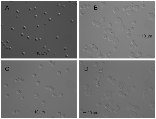
Figure 8. Magnetic separation of HSC cultures results in the improved morphology of the cells.
The morphology of cells by differential interefence contrast (DIC) microscopy. (A) The donor-derived RBCs as a control and the HSC culture before (B) and after separation: (C) “positive” fraction; (D) “negative” fraction. Note enrichment in smaller, denser cells in (C) consistent with the presence of maturing RBCs in the magnetically separated “positive” fraction. (Their size and lack of evidence of the biconcave disk morphology suggests that these are reticulocytes rather than fully mature RBCs.).

