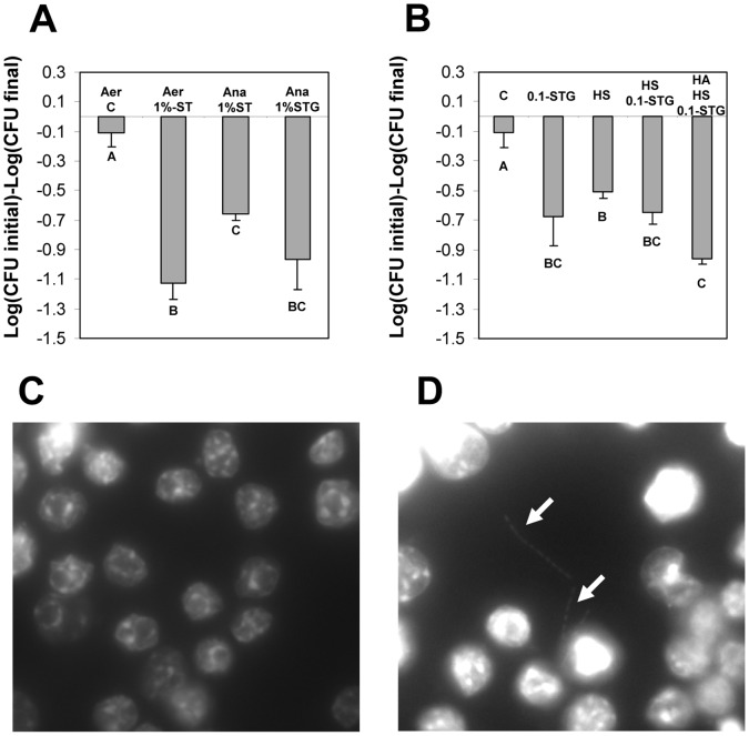
Figure 8. Factors affecting survival of C. difficile spores inside Raw 264.7 cells.
A) Monolayers of Raw 264.7 cells were infected at an MOI of 10 with dormant C. difficile spores; and with spores incubated for 30 min with 1% sodium taurocholate (ST) in DMEM and 1% sodium taurocholate-5 mM L-glycine (STG) in DMEM. After 30 min of infection, unbound spores were washed, and Raw 264.7 cells were incubated in DMEM with no FBS under: Aer, aerobic conditions-5% CO2 for 24 h; Ana, anaerobic conditions for 24 h. Loss of spore viability was determined as described in Methods section. B) Monolayers of Raw 264.7 cells were infected with non-heat activated C. difficile spores treated for 30 min with: C, DMEM; 0.1-STG, 0.1% sodium taurocholate-5 mM L-glycine in DMEM; HS, 50% human serum in DMEM; HS 0.1-STG, 0.1% sodium taurocholate-5 mM L-glycine-50% human serum in DMEM; HA HS 0.1-STG, with heat activated C. difficile spores treated for 30 min with 0.1% sodium taurocholate-5 mM L-glycine-50% human serum in DMEM. Infected monolayers of Raw 264.7 cells were incubated for 24 h and viable spores were determined by plating aliquots of lysed Raw 264.7 cells onto BHIS agar plates as described in Methods section. Treatment C corresponds to data from Fig. 7A and is shown for comparative purposes. C,D) Fluorescence micrographs of Raw 264.7 cells infected with 1.0%-STG C. difficile spores. Infected Raw 264.7 cells were incubated for 24 h under aerobic (panel C) and anaerobic (panel D) conditions, fixed and DNA material of either Raw 264.7 cells and C. difficile vegetative cells were stained with DAPI as described in Methods section. White arrows denote growing C. difficile vegetative cells. Results are the average of at least three independent experiments and error bars are standard error of the mean.

