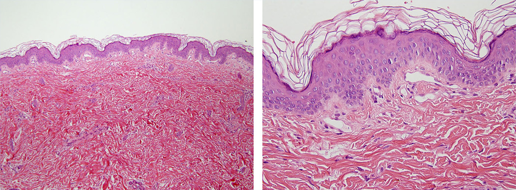
FIG. 5.
Skin biopsy images of Patient 3 (LR10-168). Routine hematoxylin and eosin stained slides at 100× and 400× magnification show a punch biopsy of skin with an unremarkable epidermis and reticular dermis. The capillaries in the papillary dermis are increased in number and are dilated as compared to normal. These biopsy findings are diagnostic of capillary malformations.

