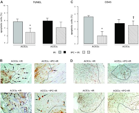
Figure 5.
Assessment of myocardial apoptosis and inflammation in ACE2c or ACE3c mice. Mice were studied after ischemia and a 24-h reperfusion period without IPC (IR) or with IPC (IPC+IR). A) Quantification of TUNEL-positive cardiomyocytes (%) in the border zone surrounding the myocardial infarction. Data are means ± se. *P < 0.05 vs. corresponding IR. B) Representative sections of hearts from ACE2c or ACE3c IR and IPC+IR mice showing the variable density of apoptic nuclei (arrows) in myocytes in the border zone. Black line delineates the infarcted area. TUNEL, original view ×40. C) Semiquantification of CD45-positive cells (score) in the infarcted area. Data are means ± se. *P < 0.05 vs. corresponding IR; †P < 0.05 vs. IPC-ACE2c. D) Representative sections of hearts from ACE2c or ACE3c IR and IPC+IR mice showing variable density of the leukocytes: inflammation is mainly limited to infarcted area. Immunohistochemistry with anti-CD45 antibody labeling all the types of leukocytes. Original view ×10.

