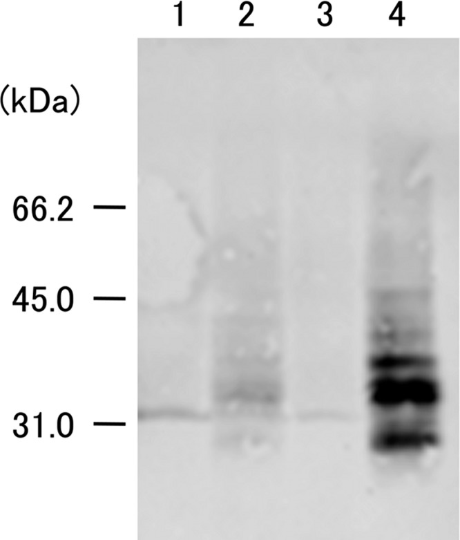
Fig 1.

Expression of poIL-18 in E. rhusiopathiae strains. Proteins in culture supernatants were precipitated with TCA, separated by SDS-PAGE, transferred onto a membrane, and probed with an anti-poIL-18 mouse MAb (11H5) (18). Lane 1, YS-1; lane 2, YS-1/IL-18; lane 3, Koganei 65-0.15; lane 4, KO/IL-18. Molecular mass markers (kilodaltons) are shown on the left.
