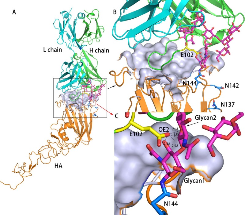
Fig 6.
Structural modeling of neutralizing antibody in complex with swine influenza virus HA antigen containing a predicted glycosylation site at N144. Panels B and C are magnified images of the antigen-antibody contact surface shown in panel A, with different magnifications focusing on different regions. The antibody-binding pocket on HA is shown using a surface diagram, while antibody and other regions of HA are shown as ribbon diagrams. Orange, HA; green, antibody heavy chain; cyan, antibody light chain. N137, N142, and N144 are colored marine (B), while glycans on N144 are colored magenta (C). In panel C, a residue on the antibody heavy chain that is close to N144 of HA is colored yellow, and the distances from the closest atoms to this residue of the heavy chain and residue N144 are shown (the atoms are antibody heavy chain E102 OE1 and HA N144 ND2). All oxygen atoms and nitrogen atoms shown are colored red and blue, respectively.

