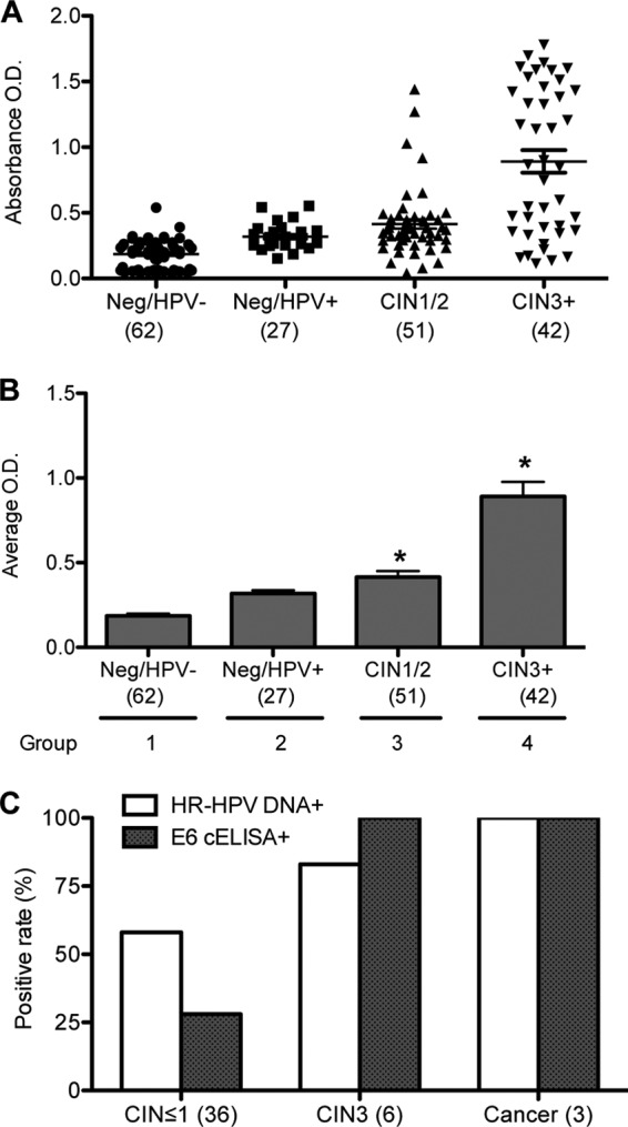
Fig 2.

Whole-cell ELISA using anti-E6 antibody. (A) Scatter dot plot of the individual absorbance signals of clinical samples in a whole-cell ELISA using anti-E6 antibody. E6 whole-cell ELISA was performed on cells from 182 cervical scrapes that were categorized into the following 4 groups: group 1, histology negative and HPV DNA negative (Neg/HPV−) (n = 62); group 2, histology negative and HPV DNA positive (Neg/HPV+) (n = 27); group 3, CIN1/2 (n = 51); and group 4, CIN3+ (n = 42). Lines show means and standard errors of the means (SEM). (B) Average absorbances obtained from the ELISA in panel A graphed as means and SEM. (C) Results from E6 whole-cell ELISA using 45 SurePath fresh samples obtained within 1 to 2 weeks of collection. The data are presented as percent positive rates for histological designations of CIN ≤ 1 (36 samples), CIN3 (6 samples), and cervical cancer (3 samples) and compared to the positive rates for HPV DNA.
