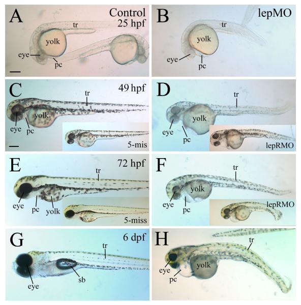
Figure 3. Gross morphological defects in leptin and leptin receptor morphants.
All images show lateral views of live embryos, with anterior to the left and dorsal up. Control embryos are shown in panels on the left column, with embryos injected with the 5-mismatched leptin MO (5- mis) shown as inserts in panels C and E. Embryos injected with the leptin MO1 (lepMO) are shown on the right column, with embryos injected with the leptin receptor MO (lepRMO) shown as inserts in panels D and F. Abbreviations: pc, pericardial cavity; sb, swim bladder; tr, body trunk. Panels A and B are of the same magnification, while remaining panels are of the same magnification as panel C. All scale bars = 200 μm.

