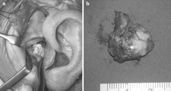
Fig. 5.
a The intra-operative photo of the patient described in case 2 showing the calcified tissue from the left TMJ. The specimen that was removed, shown in Fig. 5b, was histologically diagnosed as pseudogout. While such intra-articular pathology is rare, the patient was managed with splint therapy and medications for over 18 months before she sought a surgical opinion

