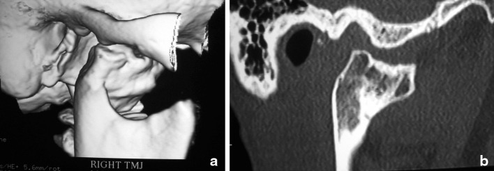
Fig. 6.
a The three dimensional CT scan of the patient described in case 3 clearly demonstrating destructive degeneration of the right TMJ which turned out to be severe posttraumatic osteoarthrosis. Figure 6b demonstrates a CT sagittal slice of the affected right condyle. The right TMJ was surgically resected and a right TMJ total joint replacement was undertaken to restore joint function and maintain the occlusion and facial symmetry

