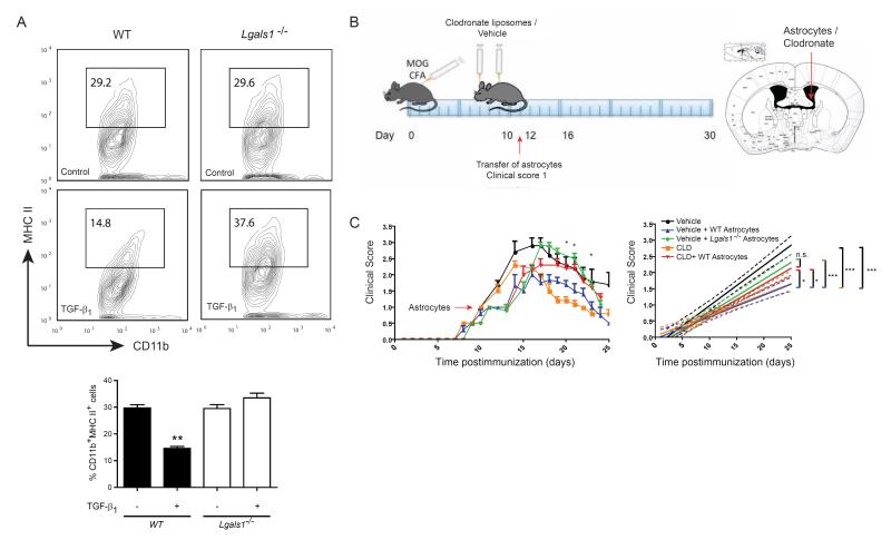
Figure 6. Astrocytes Control Microglia Activation and Limit EAE Severity via Gal1.
(A) Flow cytometry of MHC II in cultured CD11b+ microglia. Microglia were exposed to conditioned media from control or TGF-β1-stimulated WT or Lgals1−/− astrocytes. Percentage of CD11b+MHC II+ cells. (B,C) Lgals1−/− mice were immunized with 200 μg MOG35-55 and injected with PBS (vehicle) or clodronate-containing liposomes (CLD) into the right lateral ventricle (day 7 and 9 post immunization). When reaching a clinical score of 1, mice were divided into 2 groups and received either WT or Lgals1−/− (KO) astrocytes into the same injection site. (B) Diagram illustrating the experimental time line (left) and injection site (right). (C) Clinical score (left) and linear regression curves of disease (right) for each group (dashed lines, 95% confidence intervals). Data are representative (A, upper panel) or are the mean ± SEM (A lower panel, C) of three independent experiments. *P < 0.05; **P < 0.01; ***P < 0.005. See also Figure S6.

