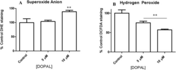
Figure 4.
Flow cytometry analysis of PC6-3 cells incubated with DOPAL for 1 h to determine levels of superoxide anion and hydrogen peroxide. DHE (A) and DCF-DA (B) were used to probe for superoxide anion and hydrogen peroxide, respectively. Cells were preincubated with each dye for 20 min, and then DOPAL was placed on cells for 1 h prior to flow cytometry analysis. All values shown represent the mean ± SEMs (n = 3). **indicates significantly more ROS production as compared to control cells where no DOPAL was present (p 0.05).

